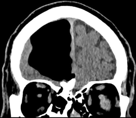
Doctors found an interesting surprise when they examined an Irish man’s brain: he seemed to be missing part of the organ.
The 84-year-old man went to the emergency department because he kept falling—a common problem among older adults—and said he had weakness on the left-side of his body, according to report published in BMJ Case Reports. Other than the weakness, which lasted for three days, medical staff noted that he didn’t have any confusion, facial weakness, vision or speech problems, and was otherwise reportedly feeling fine.
Although his blood work came back normal, brain imaging scans revealed a strange, 3.5-inch pocket of air in his head.
“We were able to see the brain scan images before receiving the formal report from our radiology specialists and immediately knew something was not right!” Dr. Finlay Brown, co-author and physician at Causeway Hospital in Northern Ireland, told Newsweek via email. “I wondered if the patient had previously undergone brain surgery or had a congenital abnormality we didn’t know about.”
But, Brown and his colleagues questioned the patient and learned he never experienced either of those.

The pocket was revealed to be a pneumatocele, a pressurized air-filled cavity, based in the brain’s right frontal lobe. Those types of cavities are typically found in patients who’ve had brain surgery or various types of infections.
Having pneumocephalus is common, but they’re typically much smaller in size.
“To find a pocket of this size in an organized fashion was extremely uncommon, with very few documented cases found while I was researching for writing up the case report,” Brown said.
The scans also revealed he had a benign bone tumor and suffered from a mini-stroke. In order to repair the air cavity, doctors presented him with the option of surgery. However, due to the associated risks, the man declined the offer. Instead, he received various types of medicines to help prevent future strokes, Brown told Live Science.
“The left-sided weakness was noted to have resolved on follow-up 12 weeks later and he remained well,” the authors conclude in the published report.
Brown hopes that his case report will encourage other physicians to fully investigate their patients, even if they present common symptoms, in order to help miss a rare cause or condition. And as for the stunning photos (seen above), those speak for themselves.
“The pictures of the scans are striking and can be easily appreciated by the general public without radiological or even medical training!” Brown said.


