
Scientists have created a groundbreaking ultrasound helmet that can target specific areas deep inside the brain without requiring surgery, the University of London announced Friday in a news release.
The device, developed by researchers from University College London and the University of Oxford, could revolutionize treatment for brain disorders like Parkinson's disease by offering a safer alternative to current surgical methods.
For years, doctors have searched for ways to treat brain conditions without cutting into patients' skulls. The new technology uses transcranial ultrasound stimulation (TUS), which sends gentle sound waves into the brain to change how nerve cells communicate with each other.
Previous ultrasound systems struggled to reach deep brain areas with enough accuracy. They often affected larger brain regions than doctors intended, making precise treatment difficult.
Participants Showed Increased Brain Activity
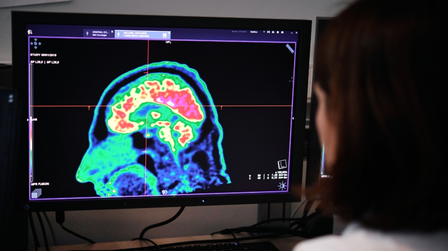
Researchers tested their device on seven volunteers, focusing on a small brain structure called the lateral geniculate nucleus (LGN). This area sits in the center of the brain and helps process visual information.During the first test, participants looked at a flashing checkerboard pattern while receiving the ultrasound treatment. Brain scans using functional magnetic resonance imaging showed increased activity in the volunteers' visual processing areas, proving the device hit its intended target.
A second experiment revealed that brain activity changes lasted at least 40 minutes after treatment stopped. This shows the system can create lasting effects on brain function.
Volunteers didn't notice any changes in their vision during the experiments, but brain scans clearly showed the neural activity had changed. Scientists hope to use such treatment to help patients with conditions like hand tremors.
Non-Surgical Treatment Potential
The technology could transform treatment for brain and mental health disorders, including Parkinson's disease, depression, and essential tremor, according to Treeby. Current treatments often require deep-brain stimulation (DBS), which involves surgically implanting electrodes in patients' brains."The ability to precisely modulate deep brain structures without surgery represents a paradigm shift in neuroscience, offering a safe, reversible, and repeatable method for both understanding brain function and developing targeted therapies," Treeby said.
The surgical option carries risks and complications, researchers emphasized. The new ultrasound system offers similar precision without requiring doctors to open patients' skulls. This could allow doctors to test brain areas for treatment potential before considering surgery or eliminate the need for operations entirely.
Several research team members have started a company called NeuroHarmonics to develop a portable version of the system. The UCL spinout company aims to make the technology available for widespread clinical use.
Dr. Eleanor Martin, the study's first author from UCL Medical Physics and Biomedical Engineering, highlighted the system's ability to work alongside brain imaging equipment.
"We designed the system to be compatible with simultaneous fMRI [functional magnetic resonance imaging], enabling us to monitor the effects of stimulation in real time," Martin said. "This opens up exciting possibilities for closed-loop neuromodulation and personalised therapies."
She said the possibility of such real-time monitoring means doctors could watch brain activity change during treatment and adjust the therapy accordingly for each patient.
Dr. Ioana Grigoras, also a first author from Oxford's Nuffield Department of Clinical Neurosciences, said the breakthrough is especially exciting for some diseases with deep brain regions that are hard to reach.
"This novel brain stimulation device represents a breakthrough in our ability to precisely target deep brain structures that were previously impossible to reach non-invasively," Grigoras said. "We are particularly excited about its potential clinical applications for neurological disorders like Parkinson's disease, where deep brain regions are especially affected."
Researchers said that more studies are needed to fully understand how ultrasound stimulation changes brain function, but noted that the results showed a major step forward in developing safe and effective brain treatment technologies.